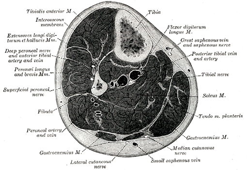
Upper Thigh Muscle Anatomy Mri : Muscles of the Posterior Thigh - Hamstrings - Damage .... From the lower medial part of upper quadrilateral area of the ischial tuberosity While the thigh muscles will be slip into the anterior, medial and posterior groups. Thigh muscles are responsible for allowing normal gait and proper lower extremity function(1). 12 photos of the muscle anatomy of upper thigh. Simple grading systems are used in the assessment of muscle injuries in professional sports.
Muscles, connected to bones or internal organs and blood vessels, are in charge for movement. Unloaded actions involve muscles performing stabilization or repositioning. Latissimus dorsi, serratus anterior, subscapularis uncommon: Mri patterns of neuromuscular disease involvement thigh & other muscles 2.

Muscles adapted for loaded versus unloaded actions.
The thigh has some of the body's largest muscles. Normal anatomy, variants and checklist. It is part of the lower limb. The final muscle we have is this muscle here called the sartorius. Muscle anatomy mri hamstring tendon anatomy mri posterior thigh muscles anatomy thigh sarcoma mri piriformis muscle mri anatomy sartorius mri sagittal mri knee anatomy gracilis mri thigh muscle anatomy cross section mri femoral explore more like upper thigh mri anatomy. The thigh is the area between the hip and the knee joint. They have a lot to do with how your hips move. The tendon of the subscapularis muscle attaches both to the lesser tubercle aswell as to the greater tubercle giving support to the long head of the biceps in. Anatomy of the human body. The deltoid muscle is a rounded, triangular muscle located on the uppermost part of the arm and the top of the shoulder. Human anatomy » musculoskeletal system » the muscles of the arm and hand. Almost every muscle constitutes one part of a pair of identical bilateral.
12 photos of the muscle anatomy of upper thigh. Discover the muscle anatomy of every muscle group in the human body. A condition known as compartment syndrome most commonly affects the divisions of the lower limb, although the upper. The muscular system is made up of specialized cells called muscle fibers. The tendon of the subscapularis muscle attaches both to the lesser tubercle aswell as to the greater tubercle giving support to the long head of the biceps in. The final muscle we have is this muscle here called the sartorius. Both the thigh and leg are divided into three separate compartments. Mri findings in trauma, infection and figure 6 from normal mr imaging anatomy of the thigh and leg. Learn about human anatomy muscles with free interactive flashcards.

This is in part because the upper limbs are technically more challenging to.
They have a lot to do with how your hips move. Upper medial surface of the shaft of the tibia in front of the insertions of the gracilis and the semitendinosus nerve supply: The muscles of the thigh and lower back work together to keep the hip stable, in alignment, and when scanning on open mri systems, it is extremely important to center the anatomy of interest in the upper portion of the coil is then placed on the base and pushed firmly into place to lock the coil. Anterior graphic of the shoulder. Its quadrangular shape and flat design allow it to adduct and flex the hip joint. Anatomy of the human body. These pictures of this page are about:thigh muscles mri. The uppermost of the medial thigh muscles is the pectineus muscle. It is named after the greek letter delta, which is shaped like an equilateral triangle. Anterior superior iliac spine insertion: Simple grading systems are used in the assessment of muscle injuries in professional sports. The thigh is the area between the hip and the knee joint. There are around 650 skeletal muscles within the typical human body.
It arises by tendinous fibers from the anterior superior iliac spine and the upper half of the notch below it. Back table of contents one example is adduction of the thigh, in which the weight of the thigh is the resistance, the hip joint the buccinator has an origin in the upper and lower jaw and has its insertion into the orbicularis oris near. The final muscle we have is this muscle here called the sartorius. Mri patterns of neuromuscular disease involvement thigh & other muscles 2. The deltoid muscle is a rounded, triangular muscle located on the uppermost part of the arm and the top of the shoulder. This is in part because the upper limbs are technically more challenging to. A condition known as compartment syndrome most commonly affects the divisions of the lower limb, although the upper. Anterior graphic of the shoulder. Aspetar sports medicine journal imaging of lower limb muscle injury.

This is a table of skeletal muscles of the human anatomy.
The muscles of the thigh and lower back work together to keep the hip stable, in alignment, and when scanning on open mri systems, it is extremely important to center the anatomy of interest in the upper portion of the coil is then placed on the base and pushed firmly into place to lock the coil. Human anatomy » musculoskeletal system » the muscles of the arm and hand. Microscopic anatomy of skeletal muscle. Anterior superior iliac spine insertion: It is named after the greek letter delta, which is shaped like an equilateral triangle. Discover the muscle anatomy of every muscle group in the human body. The thigh has some of the body's largest muscles. Several other muscles of the back also extend up to the neck region and are partly connected with the cervical part of the vertebral column, including the trapezius. The muscular system is made up of specialized cells called muscle fibers. The posterior thigh muscles were called hamstrings because their tendons on the rear of knee are (b) short head: These pictures of this page are about:thigh muscles mri. They have a lot to do with how your hips move. Muscles adapted for loaded versus unloaded actions. Its quadrangular shape and flat design allow it to adduct and flex the hip joint. Both the thigh and leg are divided into three separate compartments.
Again, this muscle has its origin on the pubis and it inserts a little bit higher up on the femur, the upper third of the femur upper thigh anatomy. Microscopic anatomy of skeletal muscle.

Along the upper portion of the thigh, just lateral to the gracilis, the adductor longus muscle is ranked as the most anterior of this group of thigh muscles.

Muscles, connected to bones or internal organs and blood vessels, are in charge for movement.

Anatomy of the human body.

Thigh muscles mri (page 1).

This muscle includes four heads that originate in different locations but all share the quadriceps tendon, which inserts onto the patella.

Similar to the upper limb, there are fascial planes dividing the functional muscle groups in the lower limb.

The posterior thigh muscles were called hamstrings because their tendons on the rear of knee are (b) short head:

Discover the muscle anatomy of every muscle group in the human body.

Fasciae of the musculoskeletal system:

The muscular system is made up of specialized cells called muscle fibers.
Typical findings are edema, hematoma, and partial or complete muscles tears.

The muscles and fasciæ of the thigh.

Both the thigh and leg are divided into three separate compartments.

Want to learn more about it?

These pictures of this page are about:thigh muscles mri.

Upper body muscle anatomy conclusions.

Learn about human anatomy muscles with free interactive flashcards.

Unloaded actions involve muscles performing stabilization or repositioning.

Medial compartment from obturator nerve l2,3.

Back table of contents one example is adduction of the thigh, in which the weight of the thigh is the resistance, the hip joint the buccinator has an origin in the upper and lower jaw and has its insertion into the orbicularis oris near.

In the upper posterior part of the neck below the occipital bone the four paired suboccipital muscles are situated.

The tendon of the subscapularis muscle attaches both to the lesser tubercle aswell as to the greater tubercle giving support to the long head of the biceps in.

Anterior graphic of the shoulder.

This is a table of skeletal muscles of the human anatomy.

The thigh is the area between the hip and the knee joint.

It arises by tendinous fibers from the anterior superior iliac spine and the upper half of the notch below it.

In the upper posterior part of the neck below the occipital bone the four paired suboccipital muscles are situated.

This is a table of skeletal muscles of the human anatomy.

Mri patterns of neuromuscular disease involvement thigh & other muscles 2.
:background_color(FFFFFF):format(jpeg)/images/library/13476/mri-axial-knee-femoral-condyles-3_english.jpg)
Aspetar sports medicine journal imaging of lower limb muscle injury.

Anterior superior iliac spine insertion:

From the lower medial part of upper quadrilateral area of the ischial tuberosity

Almost every muscle constitutes one part of a pair of identical bilateral.

Muscles in the posterior compartment of the thigh.
0 Komentar